

Introduction
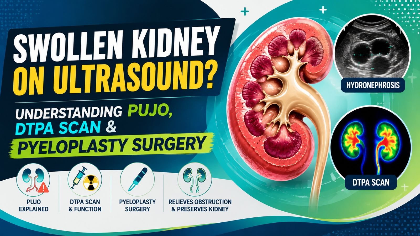
Many patients in Jaipur and across Rajasthan undergo a routine ultrasound and are told their kidney is “swollen” or shows hydronephrosis. The first assumption is almost always a kidney stone. However, not all kidney swelling is caused by stones.
One important — and often missed — condition that causes kidney swelling is Pelvi-Ureteric Junction Obstruction (PUJO). If left undiagnosed, PUJO can gradually damage kidney function over months or years, often without obvious symptoms.
This guide explains what PUJO is, why a DTPA renal scan is essential for diagnosis, and when pyeloplasty surgery becomes necessary.
What Is PUJO (PUJ Obstruction)?
PUJO occurs when the junction connecting the kidney (renal pelvis) to the ureter becomes narrowed or blocked. Since urine cannot drain normally from the kidney into the ureter and bladder:
- The kidney becomes progressively swollen
- Pressure builds up inside the collecting system of the kidney
- Kidney function may decline gradually if the blockage is not addressed
Because it can develop silently, many patients only discover it during an unrelated abdominal ultrasound.
Symptoms of PUJO
Some patients remain completely asymptomatic and are diagnosed incidentally during a health check-up or scan done for another reason.
Others may notice:
- Flank pain (pain in the side of the back, below the ribs)
- Recurrent abdominal pain
- Nausea and vomiting during episodes of pain
- Recurrent urinary tract infections (UTIs)
- Formation of kidney stones
- Intermittent pain that worsens after drinking large amounts of water (a classic clue for PUJO)
If you or a family member in Jaipur has experienced recurring one-sided flank pain that intensifies after fluid intake, this should be evaluated by a urologist rather than dismissed as generic back pain.
Primary vs Secondary PUJO
Primary PUJO
This form is present from birth (congenital) and is the most common type. Common underlying causes include:
- Congenital narrowing of the PUJ segment
- Abnormal muscle/tissue development at the junction, affecting normal peristalsis
- An abnormal crossing blood vessel compressing the junction from outside
Secondary PUJO
This develops later in life due to acquired causes such as:
- Previous kidney stone disease
- Recurrent kidney infections
- Prior abdominal or urological surgery
- Scar tissue (fibrosis) formation at the junction
- Trauma to the kidney/ureter region
Treatment approach depends on identifying which type is present and what is driving the obstruction.
Why Ultrasound Alone Is Not Enough
Ultrasound is an excellent first screening tool — it can clearly show that a kidney is swollen (hydronephrosis). However, ultrasound alone cannot reliably tell us:
- Whether the obstruction is functionally significant
- Whether the kidney is actively losing function
- Whether surgery is actually required
This is exactly why a DTPA Renal Scan is the next essential step once hydronephrosis is detected, rather than jumping straight to surgery — or, equally important, wrongly reassuring a patient that “it’s nothing” without functional testing.
What Is a DTPA Scan?
A DTPA (Diethylene Triamine Pentaacetic Acid) Renogram is a nuclear medicine scan that assesses two critical things:
- Kidney Function — how much each kidney is contributing to overall kidney function
- Urine Drainage — how efficiently urine is draining out of the affected kidney
This test is available at major diagnostic and nuclear medicine centers in Jaipur and is a standard part of PUJO evaluation.
Understanding Split Renal Function (SRF)
The DTPA scan reports what is called Split Renal Function, which compares how much each kidney is working relative to the other.
A healthy person typically shows:
- Right kidney ≈ 50%
- Left kidney ≈ 50%
Small variations within a normal range are not a cause for concern.
Example — Normal: Right kidney = 52%, Left kidney = 48%
Example — Abnormal: Right kidney = 70%, Left kidney = 30%
A significant imbalance like this may indicate that the obstructed kidney is progressively losing function, which strengthens the case for intervention.
When Is Surgery Usually Recommended?
The decision to operate is not based on swelling alone. Several factors are weighed together:
Declining Kidney Function A drop in split renal function on repeat DTPA scans over time is one of the strongest indicators for surgery.
Poor Drainage Pattern Persistent obstructive pattern seen on the DTPA drainage curve.
Symptoms Ongoing pain, recurrent infections, or recurrent stone formation in the affected kidney.
Progressive Hydronephrosis Worsening kidney swelling seen on serial ultrasound or CT imaging.
What Is Pyeloplasty?
Pyeloplasty is the definitive surgical treatment for PUJO. During this reconstructive procedure, the narrowed or obstructed segment is removed, and the healthy ureter is reconnected to the kidney’s collecting system (renal pelvis) to restore normal drainage.
The goals of pyeloplasty are to:
- Improve urine drainage from the kidney
- Preserve remaining kidney function
- Relieve pain and other symptoms
- Prevent further, irreversible kidney damage
How Is Pyeloplasty Performed?
Laparoscopic Pyeloplasty
Performed through small keyhole incisions. Benefits include:
- Less post-operative pain
- Smaller scars
- Faster recovery compared to open surgery
Robotic Pyeloplasty
Available at select advanced centers. Advantages include:
- Enhanced surgical precision
- More accurate suturing of the reconstructed junction
- Faster recovery in appropriately selected patients
Patients in Jaipur can discuss with their treating urologist which approach is most suitable based on kidney anatomy, prior surgeries, and available surgical infrastructure.
What Happens During Surgery?
- The surgeon identifies the site and cause of obstruction.
- The narrowed segment of the PUJ is removed.
- The junction is surgically reconstructed.
- A temporary DJ (Double-J) stent is placed to support healing and drainage.
- Free flow of urine from the kidney is confirmed before closing.
Success Rate of Pyeloplasty
Modern laparoscopic and robotic pyeloplasty techniques achieve success rates exceeding 90–95% in experienced hands. Most patients experience:
- Resolution of flank pain
- Significantly improved drainage on follow-up scans
- Stabilization, and often improvement, of kidney function
Recovery After Pyeloplasty
Hospital Stay: Usually 2–4 days, depending on the surgical approach and recovery.
DJ Stent: Typically removed after a few weeks in an outpatient procedure.
Return to Routine Activity: Most patients gradually resume normal daily activities within a few weeks, with heavy exertion restricted slightly longer.
Can Kidney Function Improve After Surgery?
This is one of the most common questions patients ask.
If surgery is performed early — while function is only mildly to moderately affected — kidney function may improve significantly after relieving the obstruction.
If poor function has been present for many years before diagnosis, the primary goal shifts to preserving remaining function and preventing further decline, rather than expecting major recovery.
This is precisely why timely diagnosis — through ultrasound followed by a DTPA scan when hydronephrosis is found — matters so much.
Local Care Context: PUJO Evaluation and Treatment in Jaipur
Patients in Jaipur and across Rajasthan have access to nuclear medicine facilities for DTPA scans and advanced laparoscopic/robotic urology care. For patients seeking government-supported treatment pathways:
- SMS Hospital, Jaipur and RUHS Hospital offer urology consultation and surgical services within the government healthcare system.
- Patients covered under Ayushman Bharat may be eligible for cashless treatment of PUJO and pyeloplasty at empanelled hospitals — eligibility should be confirmed with the hospital’s Ayushman Bharat helpdesk.
- Jan Aushadhi Kendra outlets across Jaipur provide affordable generic medications, including post-operative pain relief and antibiotics prescribed after pyeloplasty.
Patients are advised to get a proper urological evaluation, including a DTPA scan, before assuming that a “swollen kidney” requires immediate surgery or, conversely, dismissing it as insignificant.
Frequently Asked Questions (FAQs)
Is every swollen kidney due to PUJO?
No. Stones, tumors, ureteric strictures, vesicoureteral reflux, and other conditions can also cause hydronephrosis. A proper evaluation is needed to determine the exact cause.
क्या हर सूजी हुई किडनी PUJO के कारण होती है? नहीं। पथरी, ट्यूमर, यूरेटर में सिकुड़न (स्ट्रिक्चर), रिफ्लक्स और अन्य कारणों से भी किडनी में सूजन (हाइड्रोनेफ्रोसिस) हो सकती है। सही कारण जानने के लिए उचित जांच आवश्यक है।
Is a DTPA scan painful?
No. It is a simple nuclear medicine test that involves a small intravenous injection, followed by imaging over a period of time. It does not require anesthesia.
क्या DTPA स्कैन में दर्द होता है? नहीं। यह एक सामान्य न्यूक्लियर मेडिसिन जांच है जिसमें एक छोटा इंजेक्शन दिया जाता है और फिर कुछ समय तक इमेजिंग की जाती है। इसमें एनेस्थीसिया की आवश्यकता नहीं होती।
Can adults get PUJO?
Yes. Although PUJO is often congenital (present from birth), many patients are only diagnosed in adulthood, sometimes after decades of being asymptomatic.
क्या वयस्कों में भी PUJO हो सकता है? हाँ। हालाँकि PUJO अक्सर जन्म से मौजूद होता है, लेकिन कई मरीजों का निदान वयस्क होने के बाद ही होता है, कभी-कभी कई वर्षों तक बिना किसी लक्षण के।
Can PUJO cause kidney stones?
Yes. Poor urine drainage from the obstructed kidney increases the risk of stone formation, as stagnant urine allows crystals to form and grow more easily.
क्या PUJO के कारण किडनी में पथरी हो सकती है? हाँ। अवरुद्ध किडनी से यूरिन का ठीक से न निकलना पथरी बनने के खतरे को बढ़ा देता है, क्योंकि रुका हुआ यूरिन क्रिस्टल बनने और बढ़ने में मदद करता है।
Is pyeloplasty a major surgery?
It is a reconstructive urological surgery, but it is now commonly performed using minimally invasive laparoscopic or robotic techniques, which significantly reduce pain, scarring, and recovery time compared to traditional open surgery.
क्या पाइलोप्लास्टी एक बड़ी सर्जरी है? यह एक पुनर्निर्माण (reconstructive) यूरोलॉजिकल सर्जरी है, लेकिन अब इसे आमतौर पर लेप्रोस्कोपिक या रोबोटिक तकनीक से किया जाता है, जिससे दर्द, निशान और रिकवरी का समय काफी कम हो जाता है, पारंपरिक ओपन सर्जरी की तुलना में।
Key Takeaway
A swollen kidney on ultrasound is not always caused by a stone. PUJO (Pelvi-Ureteric Junction Obstruction) is an important and often treatable cause of hydronephrosis. A DTPA renal scan helps determine whether kidney function is being affected and whether surgery is truly necessary. When indicated, pyeloplasty — performed laparoscopically or robotically — offers strong long-term outcomes and helps preserve kidney function for the long run.
If you have been told your kidney is swollen on ultrasound, consult a urologist for a proper functional evaluation before deciding on the next step.
This article is written for general patient education and does not replace an in-person consultation. For evaluation of hydronephrosis, PUJO, or kidney drainage problems, please consult a qualified urologist.
About the Author: Dr. Mohit Sharma is an MCh Urology specialist trained at AIIMS, with clinical expertise in kidney stone disease, reconstructive urology, and minimally invasive urological surgery. He is the founder of UROWALA, a urology patient education and consultation platform based in Jaipur, Rajasthan.